Development of High-Throughput Light-Sheet Fluorescence Lifetime Microscopy for 3D Functional Imaging of Metabolic Pathways in Plants and Microorganisms
Authors:
Adam Bowman1,3* ([email protected]), Dara Dowlatshahi1,2, Rose Knight1,2, Nils Bode1, Soichi Wakatsuki1,2, Mark Kasevich1 (PI)
Institutions:
1Stanford University; 2SLAC National Accelerator Laboratory; 3Salk Institute for Biological Studies
Abstract
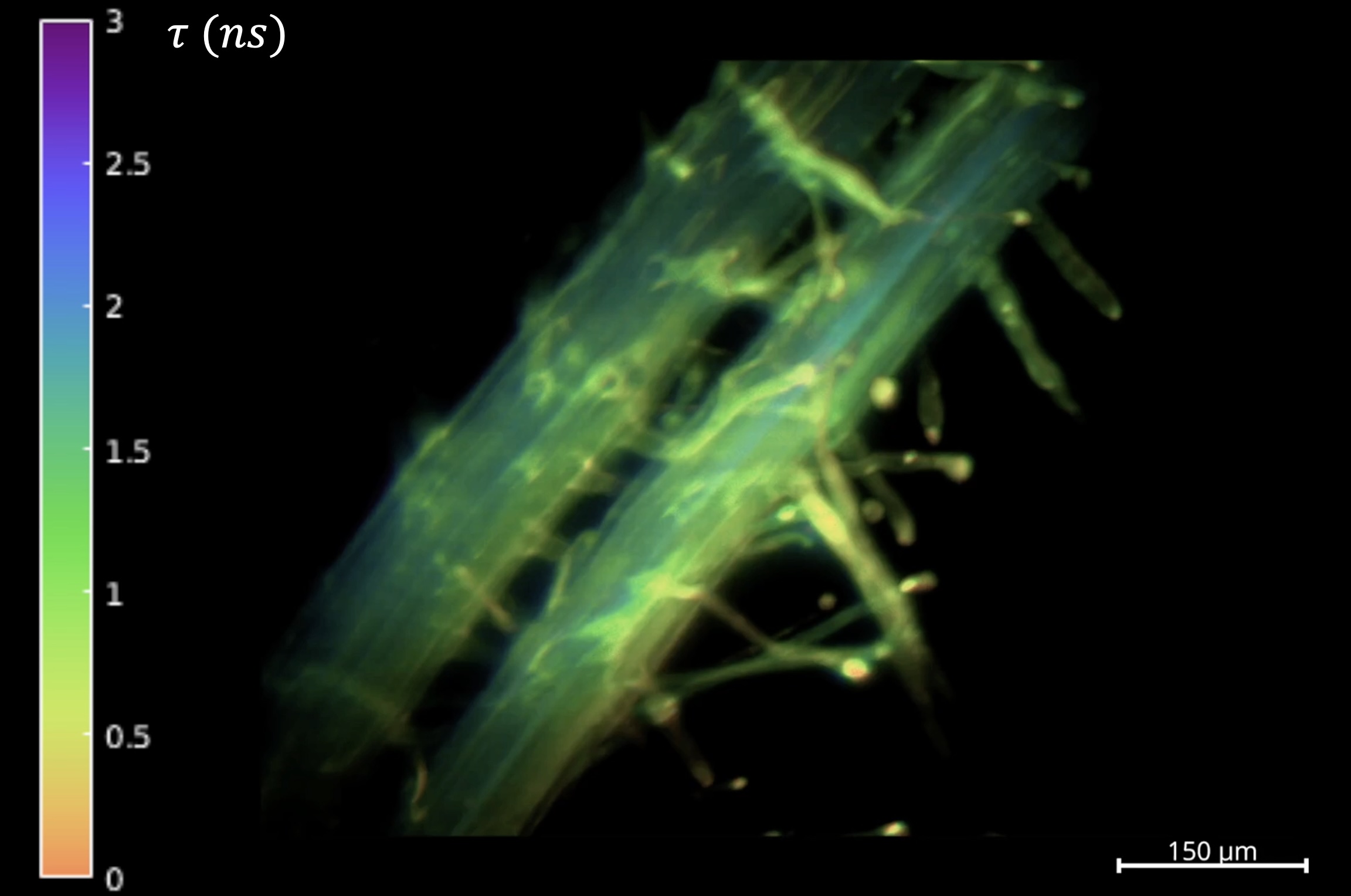
The project goal is to realize a high-speed lifetime imaging platform for light-sheet microscopy of metabolic pathways and interactions between plants and soil bacteria using electro-optic fluorescence lifetime microscopy (EO-FLIM) (Bowman et al. 2019; Bowman and Kasevich 2021). Wide-field optical modulators allow efficient lifetime capture combined with low noise readout on standard scientific cameras. This group presents results from its fluorescence lifetime light-sheet microscopy platform. Images acquired in a selective plane illumination microscope are gated using a Pockels cell driven at 80 megahertz, enabling light-sheet FLIM with up to 800 micrometer (μm) field of view. Volumetric lifetime acquisitions are demonstrated on live Arabidopsis thaliana root samples using both genetically encoded fluorescent proteins and endogenous autofluorescence. The group also presents application of EO-FLIM to record neuron activity in vivo at kilohertz frame rates using a genetically encoded voltage indicator (Bowman et al. 2023).
Image

Fluorescence Lifetime Light-Sheet Microscopy. Arabidopsis root labeled with green fluorescent protein (maximum intensity projection). [Courtesy Stanford University]
References
Bowman, A. J., and M. A. Kasevich. 2021. “Resonant Electro-Optic Imaging for Microscopy at Nanosecond Resolution,” ACS Nano 15(10), 16043–54. DOI:10.1021/ acsnano.1c04470.
Bowman, A. J., et al. 2019. “Electro-Optic Imaging Enables Efficient Wide-Field Fluorescence Lifetime Microscopy,” Nature Communications 10(1) DOI:10.1038/s41467-019-12535-5.
Bowman, A. J., et al. 2023. “Wide-Field Fluorescence Lifetime Imaging of Neuron Spiking and Subthreshold Activity In Vivo,” Science 380(6651), 1270–5. DOI:10.1126/science.adf9725.
Funding Information
This research was supported by the DOE Office of Science, through the Biomolecular Characterization and Imaging Sciences program, BER program, grant DE-SC0021976.
