BER research complementary to the Genomic Science Program
Improve or develop new multifunctional, multiscale imaging and measurement technologies that enable visualization of the spatiotemporal and functional relationships among biomolecules, cellular compartments, and higher-order organization of biological systems.
Insights into the properties, behavior, and functions of biological systems are greatly enabled by the availability of spatial, structural, and dynamic information at varying scales. Knowing how cellular components, whole cells, or populations can assemble, interact, and physically behave in time and space can provide unique clues about function and reveal opportunities for experimental manipulation.
To address the Biological and Environmental Research (BER) Program’s biomolecular characterization and imaging science goals, BER supports the development of bioimaging tools, methods, and technologies and invest in related infrastructure and resources. This support includes developing technologies for structural biology and biological imaging at subnanometer to micrometer resolution, as well as approaches for real-time, nondestructive visualization of living systems. In addition, BER seeks to satisfy unmet needs across all portfolio elements by aligning imaging and structural biology with genomic science capabilities and by leveraging DOE’s unique beamline and computational infrastructure. In light of these priorities, BER supports programs that leverage both the spatial and temporal resolutions available from neutron, photon, and electron beams, as well as the advantages offered by the direct, in situ visualization of living tissues through light, electron, and quantum science–enabled microscopy.

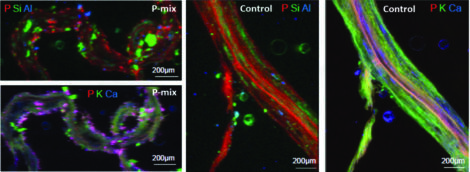
Micro-X-Ray Fluorescence (μXRF) Maps of Longitudinal Root Sections. The μXRF maps show evidence of phosphorus uptake in a plant that has the endophytic bacterial strains WP5 and WP42 (“P-mix”). The P-mix sample exhibits phosphorus hot spots, while the control displays a more homogeneous phosphorus distribution. Phosphorus appears pink in the bottom left and far right images due to phosphorus and calcium overlapping. [Reprinted under a Creative Commons Attribution License (CC BY 4.0) from Varga, T., et al. 2020. “Endophyte-Promoted Phosphorus Solubilization in Populus,”Frontiers in Plant Science 11, 567918.]
Characterizing complex systems at a range of spatial and temporal scales often requires multiple methods. Integrating methodologies to connect molecular properties to system-level functions is therefore a priority. Examples of targeted research areas include understanding how macromolecules and complexes are structured, how interactions at the macromolecular level confer function (i.e., the workings of “molecular machines”), how molecular assemblies are organized and networked in the cellular environment, and, ultimately, how their pathways are regulated to achieve functional characteristics. Integrating experimental capabilities via computational resources is critically needed to synthesize requisite measurements across multiple scales of length and time. Modern imaging technology creates enormous amounts of data that require computationally intensive processing and interpretation. These capabilities, along with the need to integrate image data with other biological characterization data, represent challenges requiring new mathematical and computational approaches. BER therefore seeks synergy with its computational biology portfolio elements and support research that meets relevant data integration challenges.
Bio- and Quantum-Imaging Technologies
BER supports fundamental research to enable novel bioimaging and characterization technologies for nondestructive, in situ, real-time measurements and their integration with omics measurements. Observing cellular components in vivo and in real time provides an indispensable perspective of biological systems in their living context. Advances in both computational and optical techniques can thus offer new insights into questions relevant to BER researchers. New technologies may, for example, enable measurements of enzyme function within cells, tracking of metabolic intermediates in vivo, monitoring of substrate and bioproduct transport within cells or across cellular membranes, and understanding of signaling processes among cells within plant-microbe and microbe-microbe interactions.
BER supports research on multifunctional technologies to image, measure, and model key metabolic processes within and among microbial cells and multicellular plant tissues. This research includes new efforts to leverage quantum-based phenomena to develop novel imaging modalities. Multiphoton microscopy has enabled higher-resolution and deep-section imaging of thick biological samples, but classical light absorption requires high laser fluence that can be damaging and cause significant perturbation for in vivo imaging. By contrast, entangled photon absorption uses much lower photon fluxes, thereby enabling new imaging modalities. Moreover, quantum-entangled imaging can be combined with quantum probes with high multiphoton cross-sections, multiple chemical functionalization and molecular tracking, spectrally tunable emission, and quantized absorption and emission states to enable high absorption of multiple entangled photons. Single-quantum emitter probes coupled with quantum-entangled photon imaging can thereby enable subdiffraction-limited imaging in vivo. Potential quantum-enabled imaging approaches may thus dramatically enhance the ability to measure biological processes in and among living cells and enable dynamic localization and imaging of cellular processes. Learn more >>>
Overall Biomolecular Characterization and Imaging Science Objectives
- Enhance the accessibility of bioimaging and structural biology infrastructure within the research community and at DOE user facilities.
- Develop and enhance tools for sample handling and transfer, optimizing the samples for multiple imaging modalities and approaches.
- Develop fast and sensitive detectors with extremely high rates of data collection and the necessary computational tools to handle large, real-time, noisy, multimodal, and multiscale data.
- Develop multifunctional, in situ, and nondestructive observation technologies for repetitive sample analyses for systems biology research.
- Visualize the spatial and temporal dynamics of expressed biomolecules within or between living plant or microbial cells and their communities.
- Explore quantum science concepts for optical imaging and sensing of cellular processes.
- Incorporate newly developed technologies into DOE user facilities or provide opportunities for commercial development through DOE programs for Small Business Innovation Research (SBIR) and Small Business Technology Transfer (STTR).
