Integrative Imaging of Plant Roots During Symbiosis with Mycorrhizal Fungi
Authors:
Andreas E. Vasdekis1* ([email protected], PI), Scott E. Baker2, Demetrios Christodoulidis3,4, Maria Harrison5, Luke Sheneman1, Ram Kasu1, Jinming Zhang1, Haokun Luo3,4
Institutions:
1University of Idaho; 2Pacific Northwest National Laboratory; 3University of Central Florida; 4University of Southern California; 5Boyce Thompson Institute
Goals
Research Plan: The goal of this project is to create an integrative optical imaging platform capable of independently quantifying the growth of plant roots and their metabolic interactions with symbiotic mycorrhizal fungi. To accomplish this, this research team is integrating interferometric (or quantitative phase) imaging with light-sheet fluorescence and Raman microscopy. This team’s approach to enable deep imaging within root tissue also involves the strategic combination of specially designed optical beams with ultralow light detection. Researchers deploy this strategy in order to overcome the challenges of the spatiotemporal degradation experienced by light as it propagates through tissue. To support these methodological advancements, group members deploy a mix of theoretical analyses, deep learning techniques for image reconstruction, and the development of dedicated biomarkers.
Abstract
Current and Anticipated Accomplishments: Since its beginning (2021), the project has concentrated on four areas. First, the team combined photon-sparse imaging with Airy light-sheet microscopy to demonstrate video Raman imaging rates at more than 1,000-fold lower irradiance than current approaches (Dunn et al. 2023; Dunn et al. 2024). This technique allowed researchers to quantify fungal metabolic using a deuterated biomarker, while the group is presently concluding its investigations on further improving these gains via alternative image reconstruction strategies (Sheneman et al. in review 2024).
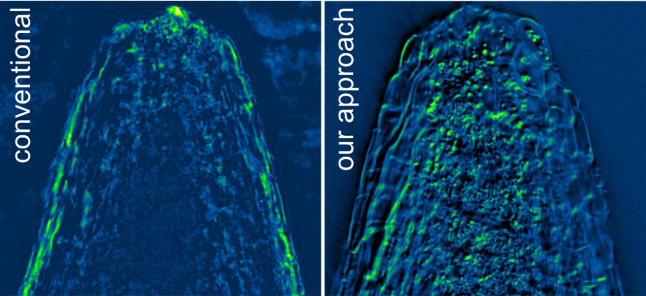
Second, the team developed a quantitative-phase microscope capable of measuring the dry density and mass within root tissue. This method integrates asymmetric illumination interferometry with standard differential interference contrast microscopes and enables the visualization of distinct features in the meristem of roots up to ~500 μm in diameter (see figure; Zhang et al. 2023 in review; Zhang and Vasdekis 2023).
Third, the group constructed a light-sheet fluorescence microscope that improves imaging efficiency in tissue via specially designed optical beams that can self-heal after scattering, and demonstrated the benefits of this technique in root tissue imaging. Fourth, the group is expanding its palette of biomarkers to track cellular changes underlying host cell accommodation of symbionts. Next steps involve: (1) enhancing the imaging depth in root tissue by refining optical fields temporally and spatially; and (2) investigate plant growth in microfluidics prior to transitioning to imaging the fungal-root symbiosis.
Benefits and Applications: This optical imaging system provides quantitative insights into root-fungi interactions, supporting the DOE’s energy prosperity goals with innovative tools, while it relies on commercial hardware and open-source software, improving its availability to the wider scientific community.
Image

Quantitative-Phase Image of a Medicago truncatula Root-Tip. Left: Using conventional methods and (Right) this team’s approach. [Adapted from Zhang, J., et al. 2024. "Quantitative Phase Imaging by Gradient Retardance Optical Microscopy," Scientific Reports 14, 9754. DOI:10.1038/s41598-024-60057-y. Republished under Creative Commons license (CC BY 4.0)]
References
Dunn, L. et al. 2023. “Video-Rate Raman-Based Metabolic Imaging by Airy Light-Sheet Illumination and Photon-Sparse Detection,” Proceedings of the National Academies of Sciences 120(9), e2210037120 (2023). DOI:10.1073/ pnas.2210037120.
Dunn, L., et al. 2024. “Video-Rate Spontaneous Raman Imaging and Method for Using.” Patent Application 18/408,198.
Sheneman, L., et al. 2024. In review. “Imaging with No Photons.”
Zhang, J., and A. E. Vasdekis. 2023. “Cost-Effective Interferometric Imaging Module Compatible with Standard Commercial Microscopes.” Provisional Patent Application 63/601582.
Zhang, J., et al. In Review. “Gradient Retardance Optical Microscopy.”
