Nondestructive, Three-Dimensional Imaging of Processes in the Rhizosphere Utilizing High-Energy Photons
Authors:
Shiva Abbaszadeh1* ([email protected], PI), Weixin Cheng1, Craig S. Levin2, Adam S. Wang2
Institutions:
1University of California–Santa Cruz; 2Stanford University
Abstract
Root–soil interactions play pivotal roles in biogeochemical processes from local scales to the global scale. The movement and transformation of nutrients, water, and soil organic matter in the root-soil interface or the rhizosphere are commonly referred to as rhizosphere processes, which include nutrient cycling, water fluxes, and carbon sequestration. Because of these crucial roles of the rhizosphere, better understanding of rhizosphere dynamics is highly needed.
To augment scientists’ capabilities to study the rhizosphere, this research group is exploring the potential of utilizing positron emission tomography (PET) and computed tomography (CT) for dynamic 3D imaging of intact rhizospheres. The CT system can provide high-resolution structural information, such as soil aggregates and pore networks, and the PET system can provide functional information at a super high temporal resolution by using a carbon-11 labeled carbon dioxide tracer (11CO2). The group has designed and developed a 11C labeling and tracing chamber inside the fume hood (130 × 60 × 80 cm3) located at the Cyclotron and Radiochemistry Facility at Stanford University.
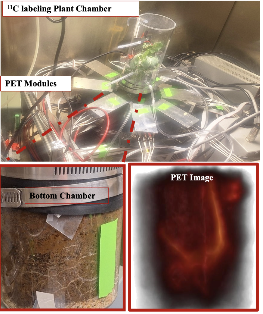
In collaboration with Jefferson Laboratory, a PET system consisting of eight PhytoPET modules was built and tested using the 11C labeling and tracing system (see figure, this page). Currently, this research team is performing image normalization and attenuation correction to understand the quantitative accuracy of the PET system. Once the PET system is ready for further experimenting, researchers will demonstrate its capability using a plant-soil system with a different soil matrix to analyze the 3D dynamic coordination between rhizospheric hotspots from PET imaging and the 3D soil matrix from CT imaging. These rhizospheric hotspots often represent a very small fraction of the soil volume but can be responsible for a substantial portion of the overall biogeochemical functions. Current methods do not have the capability to visualize these crucial hotspots inside intact soil matrix.
An amorphous selenium (a-Se) direct conversion detector on a scalable Complementary Metal-Oxide- Semiconductor (CMOS) readout for a large capability is under development at University of California–Santa Cruz for high spatial resolution CT imaging (<20 micrometer resolution). The potential capability of the proposed hybrid detector for imaging very fine soil structures at less than 20 μm resolution has been demonstrated based on a previously developed prototype [1k × 1k Readout Integrated Circuit (ROIC) by KA Imaging]. Successful development of accessible PET/CT systems for rhizosphere imaging and direct observation of belowground ecosystems can reveal the puzzling complexity and crucial Earth system functions of root–soil systems. This new capability also has important implications for other disciplines in Earth system science.
Image

11C Labeling and Tracing Chamber. Shown inside the fume hood with eight PhytoPET detector modules surrounding the bottom portion of the chamber, which is sealed/separated from the top chamber. The carbon-11 labeled carbon dioxide tracer gas is release to the top chamber containing plant canopy. The root soil is contained in the bottom chamber; the 11C activity position emission tomography image of the bottom chamber is shown, which represents active root systems and the soil in the rhizosphere network receiving root exudates. The bright curved picture on the right and the bottom portions of the image shows the tap root and bundles of fine roots. [Courtesy University of California– Santa Cruz]
