Diffraction-Free Beams in Light-Sheet Microcopy
Authors:
Haokun Luo ([email protected])1*, Ramachandran Kasu2, Andreas E. Vasdekis2, and Demetrios N. Christodoulides1
Institutions:
1University of Southern California; and 2University of Idaho
Goals
To overcome the tradeoff between frame rates and levels of irradiance in Raman imaging, the team introduces a prudently constructed light-sheet microscope relying on the Airy beam. This unique propagation-invariant beam also enhances considerably the field of view and contrast in light-sheet microscopy compared to traditional Gaussian beams. Further, the bending properties of accelerating diffraction-free beams are explored to aid the rates and resolution of light-sheet microscopy.
Abstract
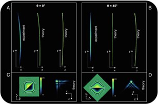
Raman imaging represents only a modest fraction of all research and clinical microscopy to date even though it exhibits great potential. This is due to the fact that most biomolecules present ultralow Raman scattering cross-sections, which impose low-light or photon-sparse conditions. Bioimaging under such conditions is suboptimal, as it either results in ultralow frame rates or requires increased levels of irradiance. Here, researchers overcome this tradeoff by introducing Raman imaging that operates at both video rates and 1,000-fold lower irradiance than state-of-the-art methods. To accomplish this, researchers deployed a judicially designed Airy light-sheet microscope to efficiently image large specimen regions (Dunn, et al. accepted, PNAS). The Airy beam is a special diffraction-free beam that harnesses its self-healing and refocusing characteristics. As such it can be utilized in light-sheet imaging (LSI) [Subedi et al. 2020, Subedi et al. 2021) including selective plane illumination imaging schemes (SPI). The diffraction-free behavior of Airy beams in these settings is investigated as a function of its cubic phase modulation ‘𝛼𝛼’, both theoretically and experimentally, as displayed in Fig. 1.
Furthermore, light-sheet microscopy facilitates wide field of view (FOV), high-contrast by employing propagation-invariant beams. To improve these properties, researchers are exploring the refocusing and self-bending properties of Airy beams and one-dimensional Bessel accelerating waves, as per Fig. 2.
Image

Fig. 1. Experimental and theoretical trajectories of the accelerating Airy beam (α = 1.09 mm−3) at two tilt angles (θ = 0° and 45° with respect the y-axis). The experimental results (xy-plane of Fig. 1B, 20× magnification, λ ~ 735 nm) represent direct Raman imaging in a PDMS slab; the theoretical trajectories present only the trajectory of the main Airy lobe in both the xy- and yz-planes. Theory and experiments were found to agree with the theoretical result in the yz-plane suggesting differences in the 3D distribution of the light at θ = 0° and θ = 45°.
References
Dunn, L., et al. Accepted. “Video-Rate, Airy Light-Sheet and Photon-Sparse Metabolic Imaging by Spontaneous Raman,” PNAS.
Subedi, N. R., et al. 2020. “Integrative Quantitative-Phase and Airy Light-Sheet Imaging,” Sci Rep 10, 20150. DOI:10.1038/s41598-020-76730-x.
Subedi, N. R., et al. 2021. “Airy Light-Sheet RamanImaging,” Opt. Express 29(20), 31941–51.
Funding Information
This research was supported by the DOE Office of Science, Office of Biological and Environmental Research (BER), grant no. DE-SC0022282.
